All diagrams in supply, synthesis and cofactors
DBH and PNMT compartment detail
DBH acts in secretory vesicles; PNMT acts in cytosol of expressing cells. These branches require the relevant enzymes; not every neuron has both.
DBH and PNMT compartment detail
Scoped educational mechanism
Open larger diagram · Full-size PNG · Diagram page
Read the diagram explanation and sources
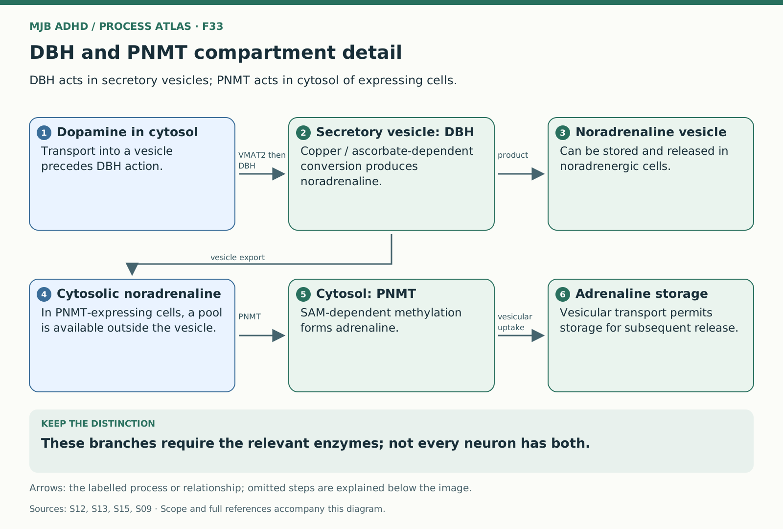
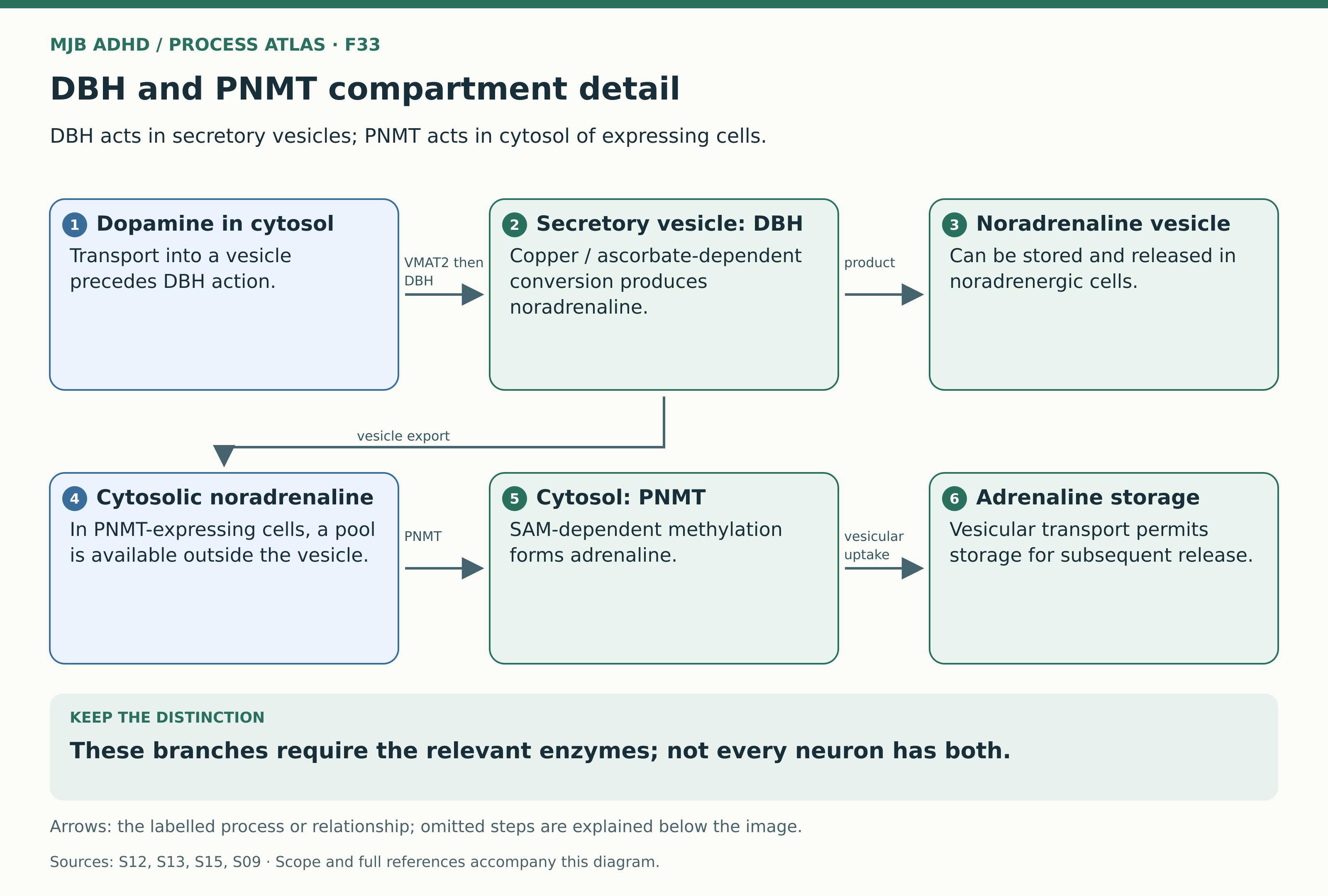
DBH is located in secretory vesicles, including noradrenergic neurons and chromaffin cells. It uses copper and ascorbate in conversion of dopamine to noradrenaline. In PNMT-expressing cells, noradrenaline is available in cytosol for SAM-dependent methylation to adrenaline, which can then be repackaged. The paired compartments correct the impression that both enzymes act freely in the same space or that every dopamine cell produces adrenaline.
- Dopamine in cytosol → Secretory vesicle: DBH: VMAT2 then DBH
- Secretory vesicle: DBH → Noradrenaline vesicle: product
- Secretory vesicle: DBH → Cytosolic noradrenaline: vesicle export
- Cytosolic noradrenaline → Cytosol: PNMT: PNMT
- Cytosol: PNMT → Adrenaline storage: vesicular uptake
What the diagram shows
DBH is located in secretory vesicles, including noradrenergic neurons and chromaffin cells. It uses copper and ascorbate in conversion of dopamine to noradrenaline. In PNMT-expressing cells, noradrenaline is available in cytosol for SAM-dependent methylation to adrenaline, which can then be repackaged. The paired compartments correct the impression that both enzymes act freely in the same space or that every dopamine cell produces adrenaline.