Process atlas
A visual way into biology: choose a topic, look at the process, then open the explanation when you need more detail.
Normal physiology, research associations and individual clinical conclusions are different kinds of information. Each diagram names its scope and links its sources. The images are educational schematics rather than diagnostic tests or personal treatment instructions.
Overview and genetic evidence
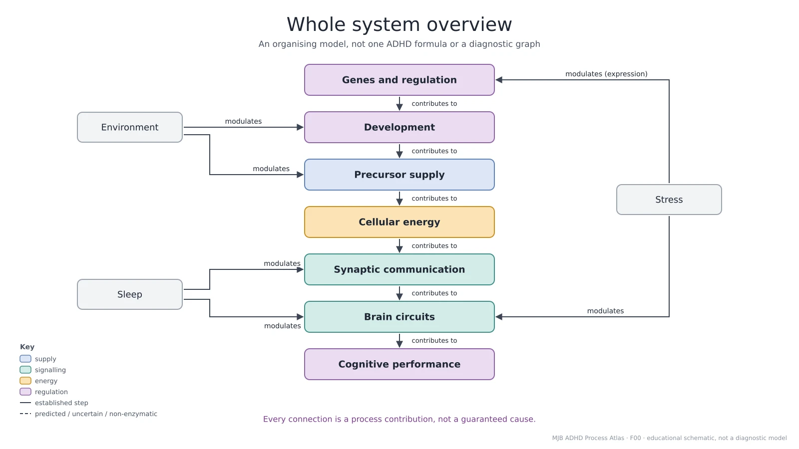 Whole system overviewAn organising framework
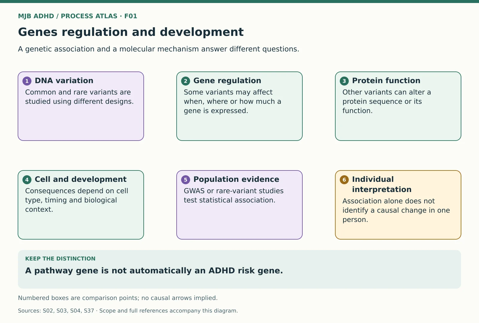 Genes regulation and developmentEvidence interpretation
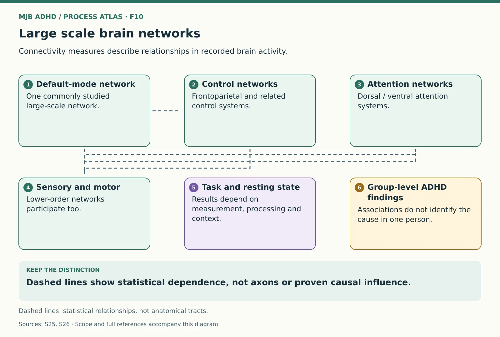 Large scale brain networksEvidence interpretation
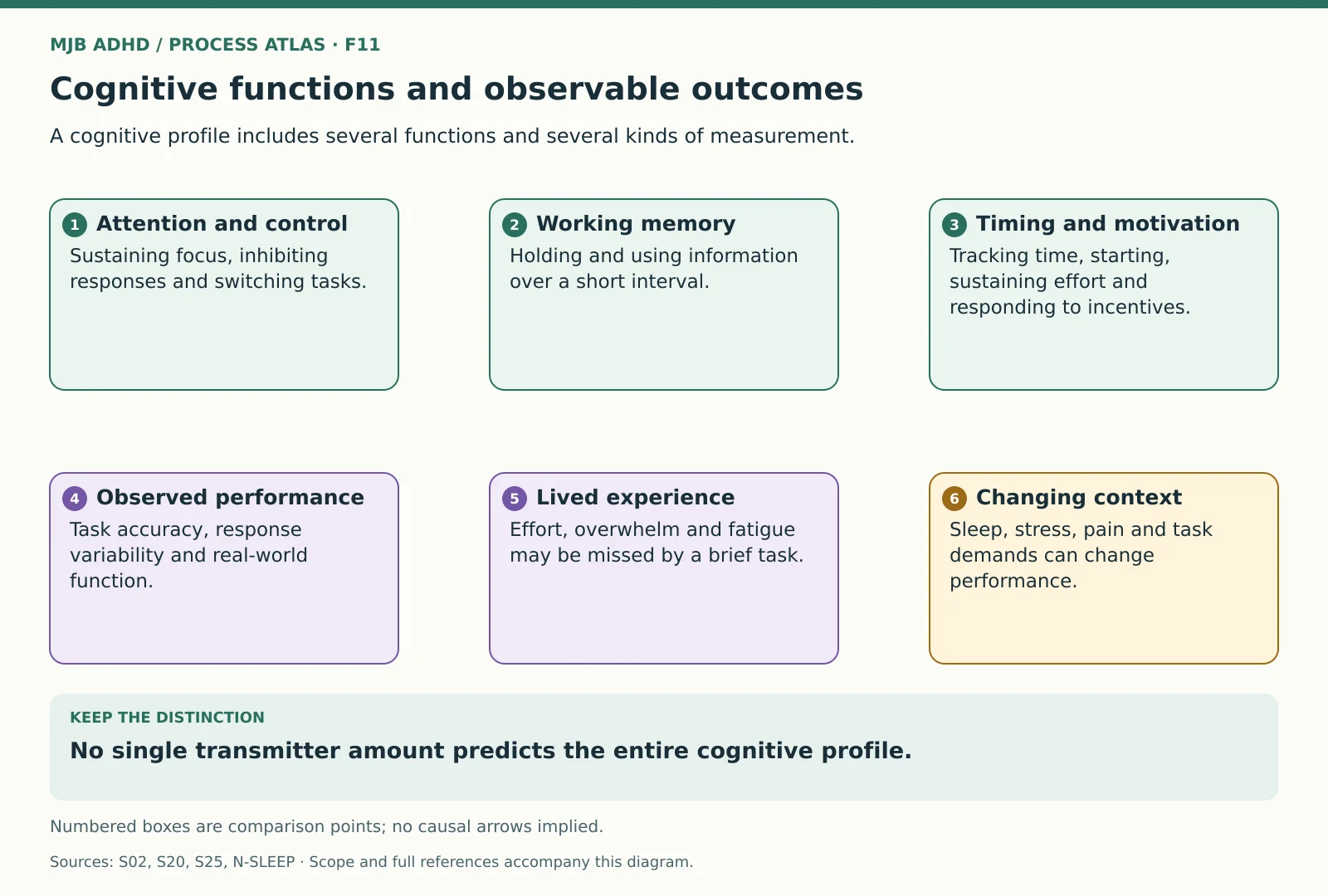 Cognitive functions and observable outcomesEvidence interpretation
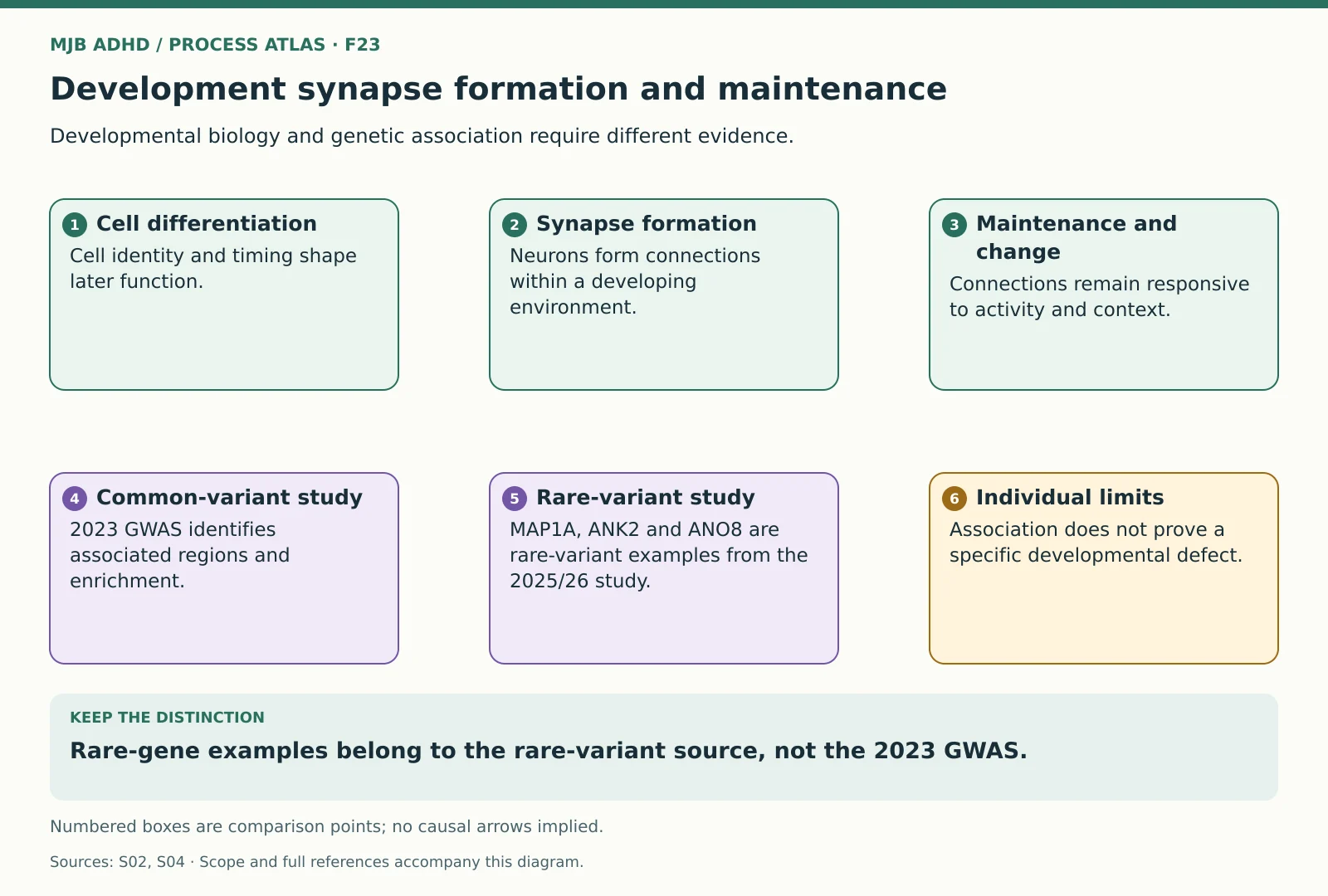 Development synapse formation and maintenanceEvidence interpretation
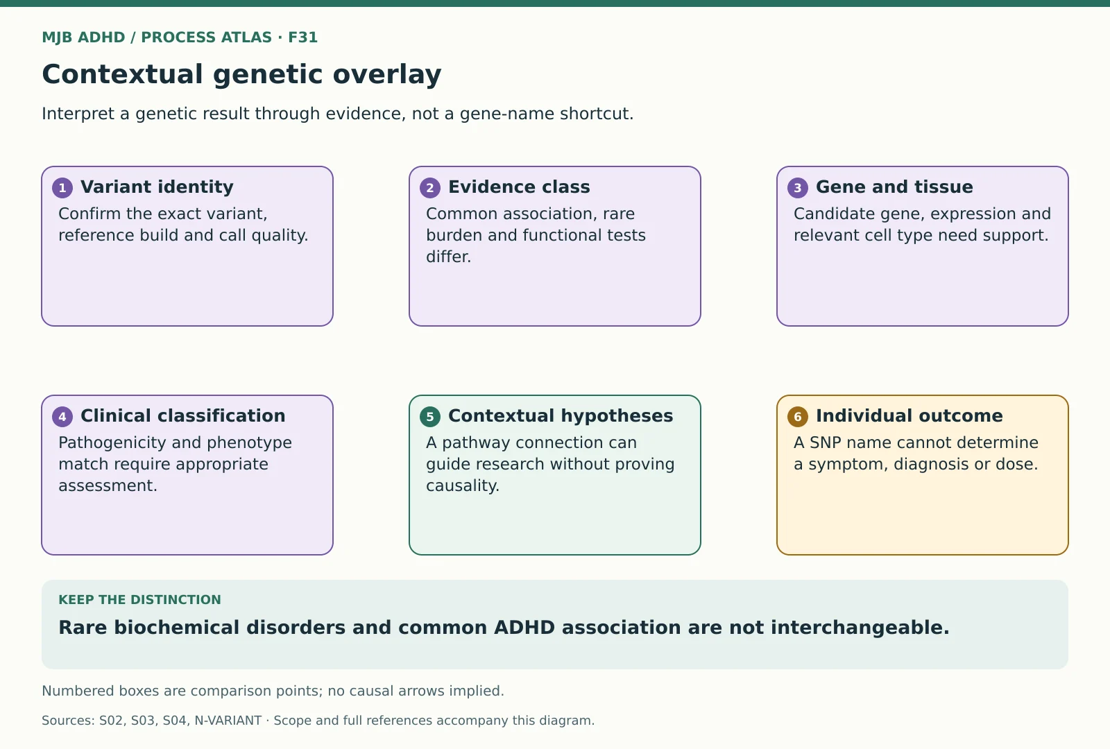 Contextual genetic overlayEvidence interpretation
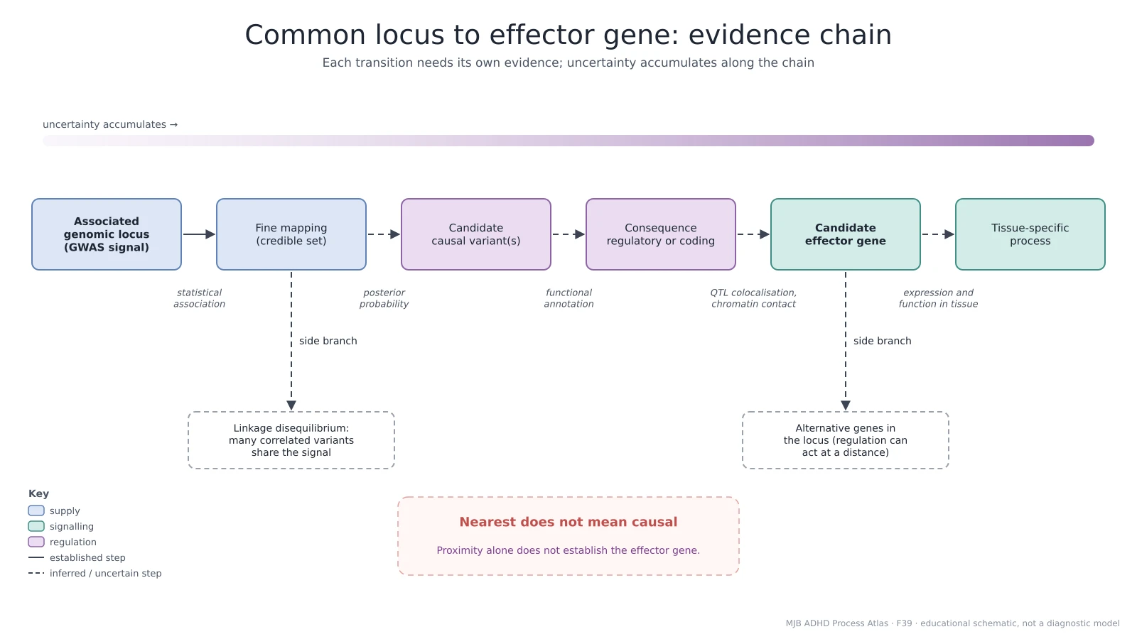 Common locus to effector gene evidenceEvidence interpretation framework
Supply, synthesis and cofactors
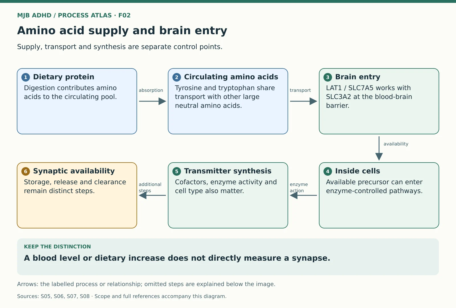 Amino acid supply and brain entryScoped educational mechanism
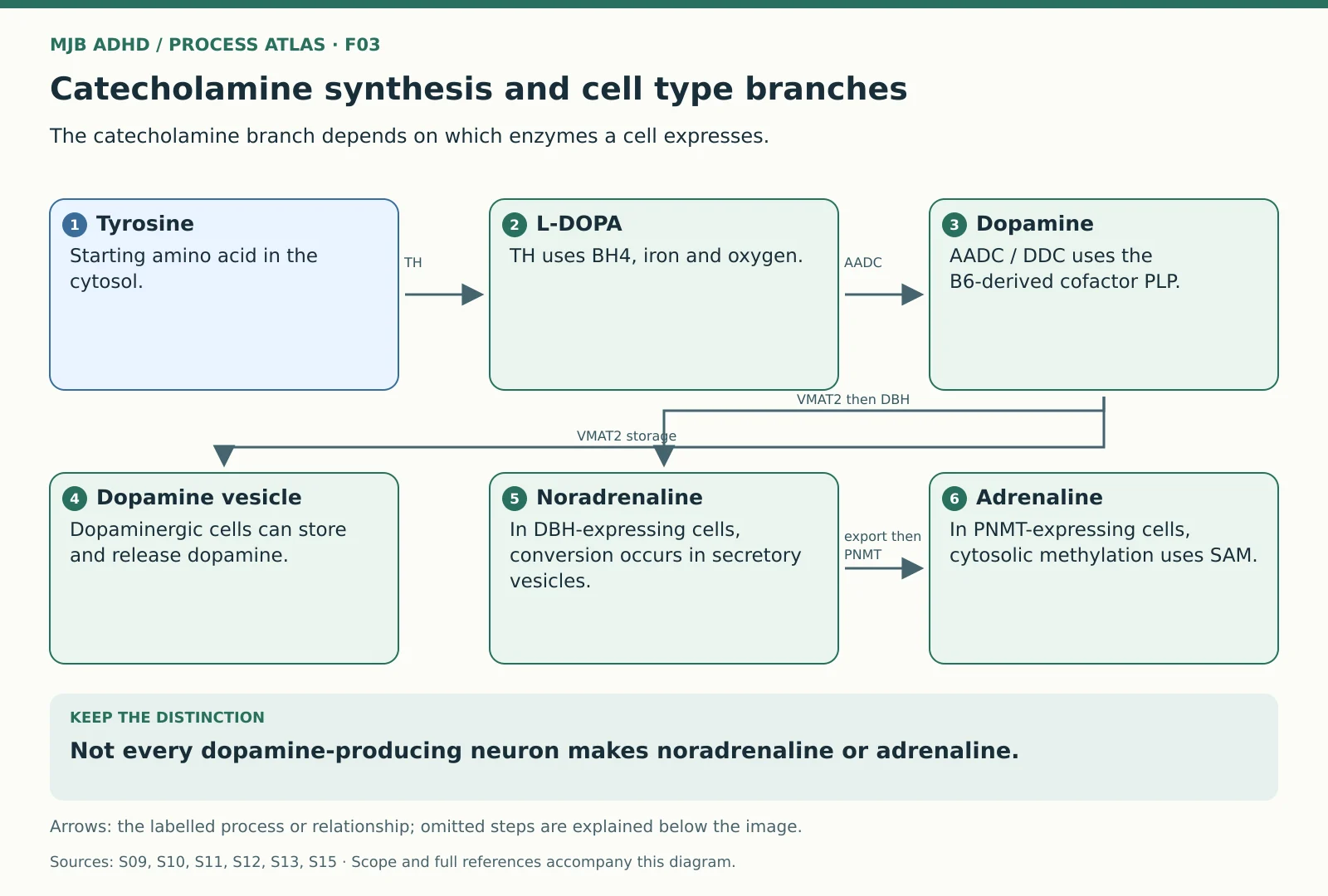 Catecholamine synthesis and cell type branchesScoped educational mechanism
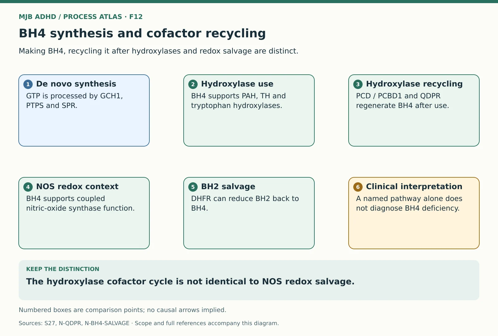 BH4 synthesis and cofactor recyclingScoped educational mechanism
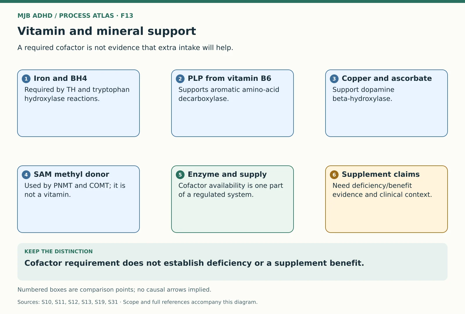 Vitamin and mineral supportScoped educational mechanism
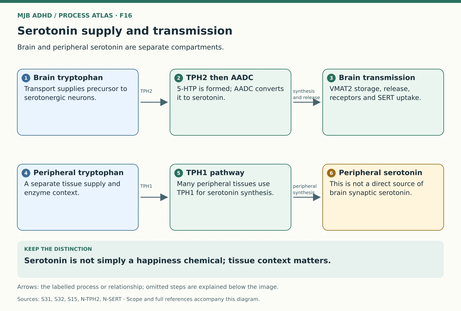 Serotonin supply and transmissionScoped educational mechanism
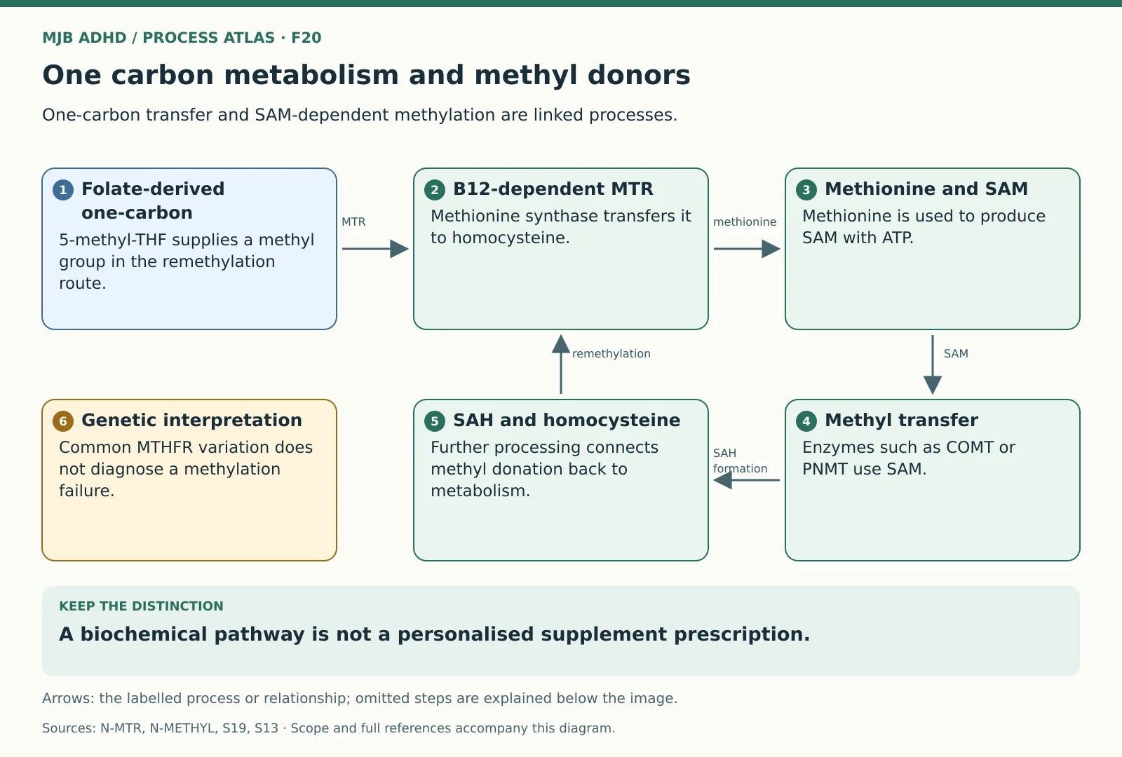 One carbon metabolism and methyl donorsScoped educational mechanism
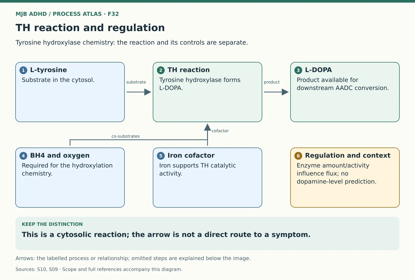 TH reaction and regulationScoped educational mechanism
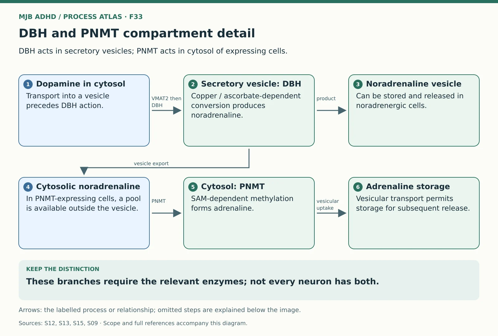 DBH and PNMT compartment detailScoped educational mechanism
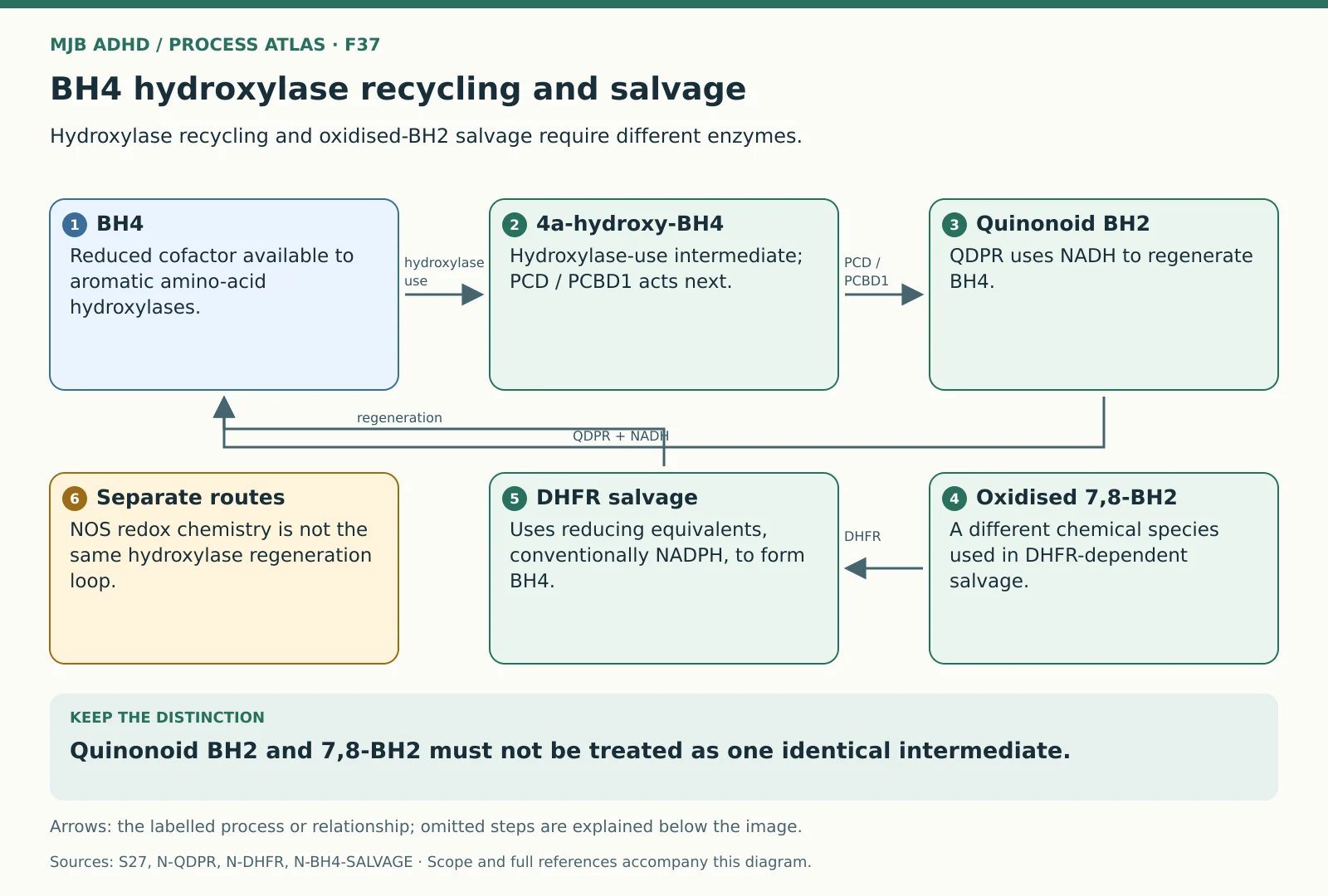 BH4 hydroxylase recycling and salvageScoped educational mechanism
Synapses and receptor signalling
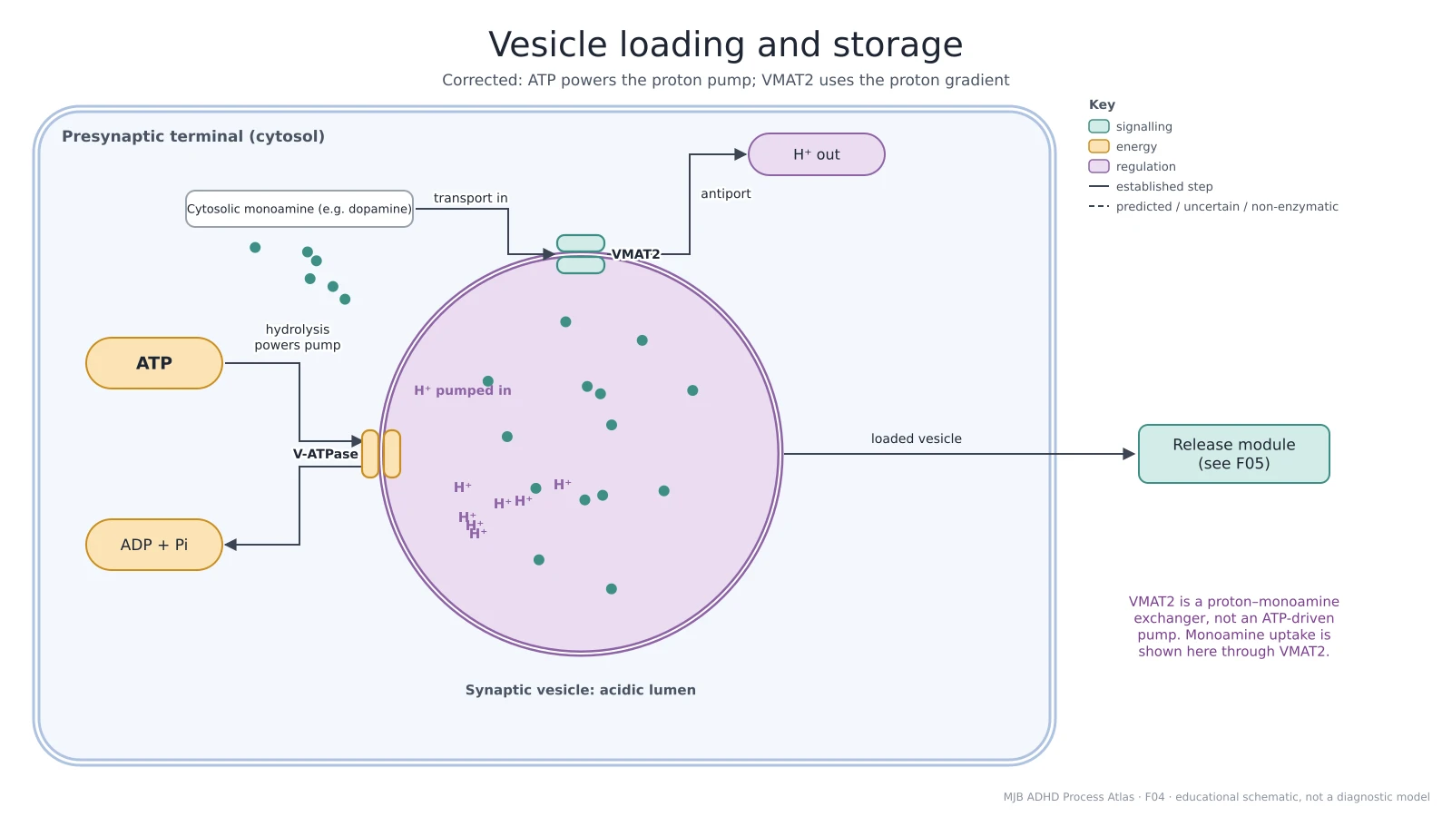 Vesicle loading and storageEstablished transport mechanism
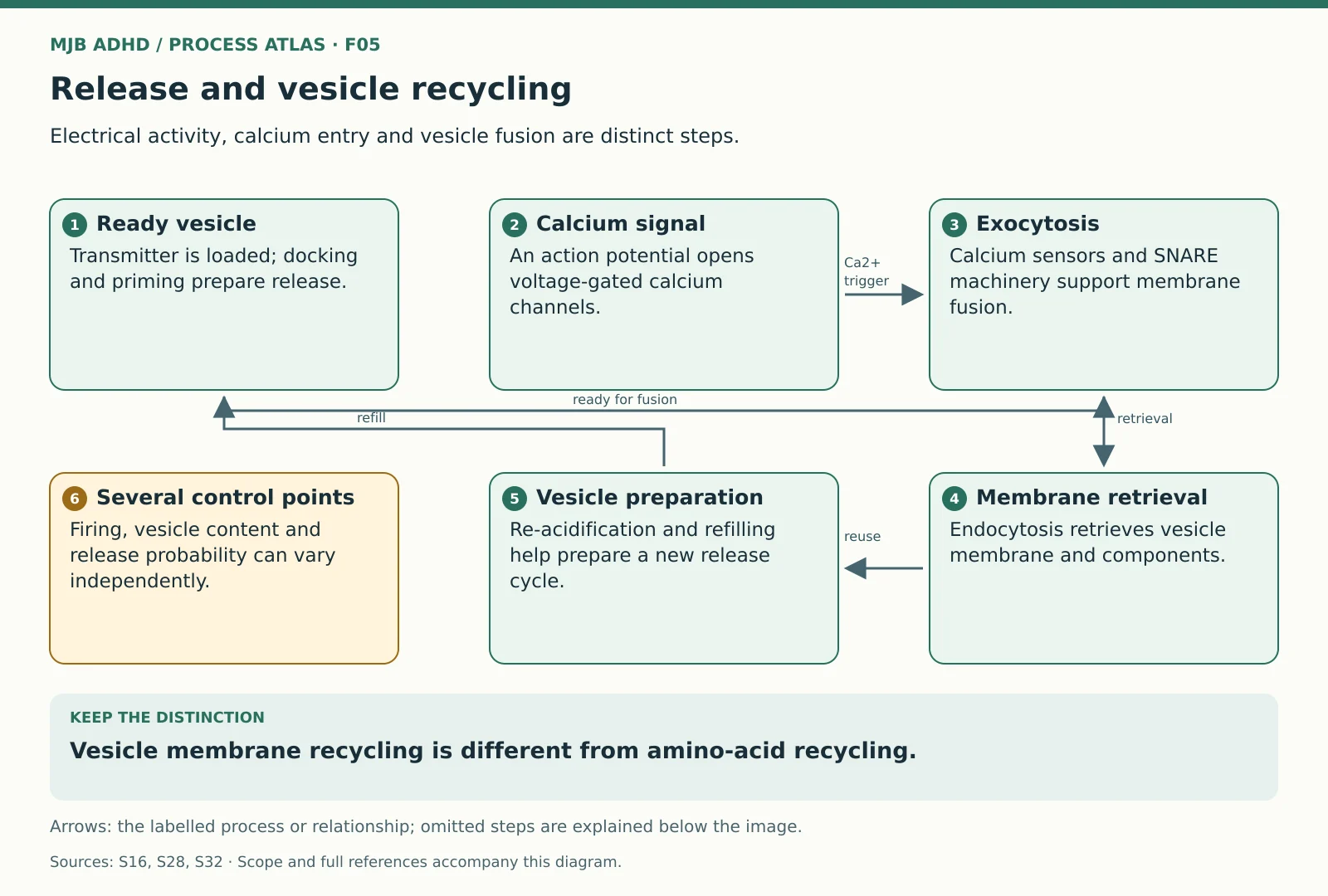 Release and vesicle recyclingScoped educational mechanism
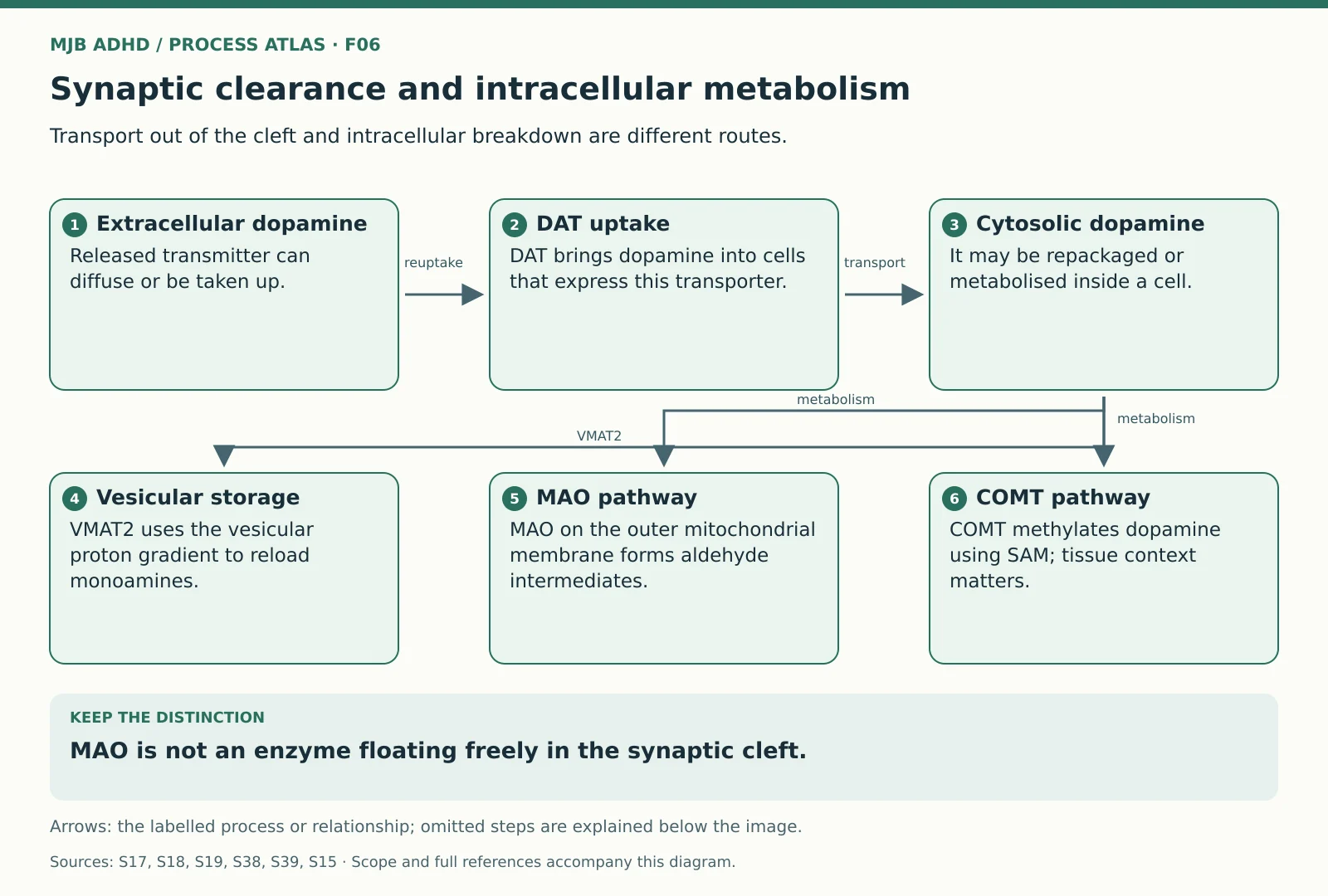 Synaptic clearance and intracellular metabolismScoped educational mechanism
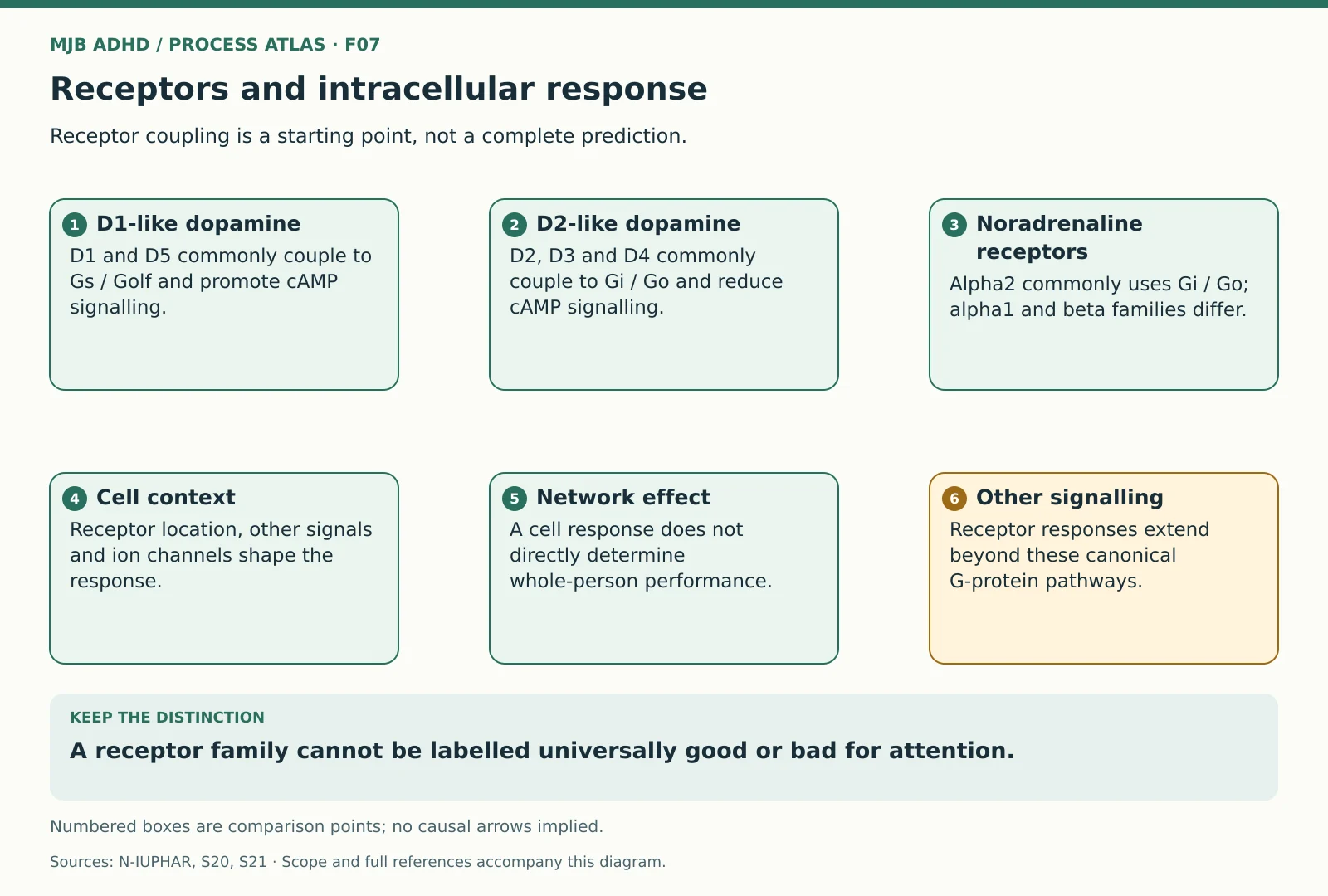 Receptors and intracellular responseScoped educational mechanism
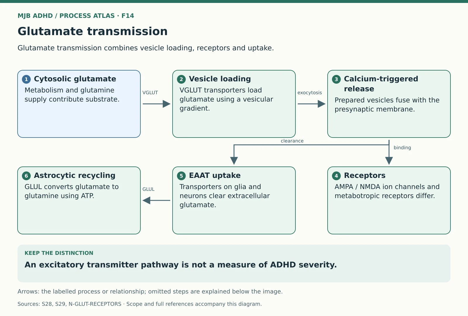 Glutamate transmissionScoped educational mechanism
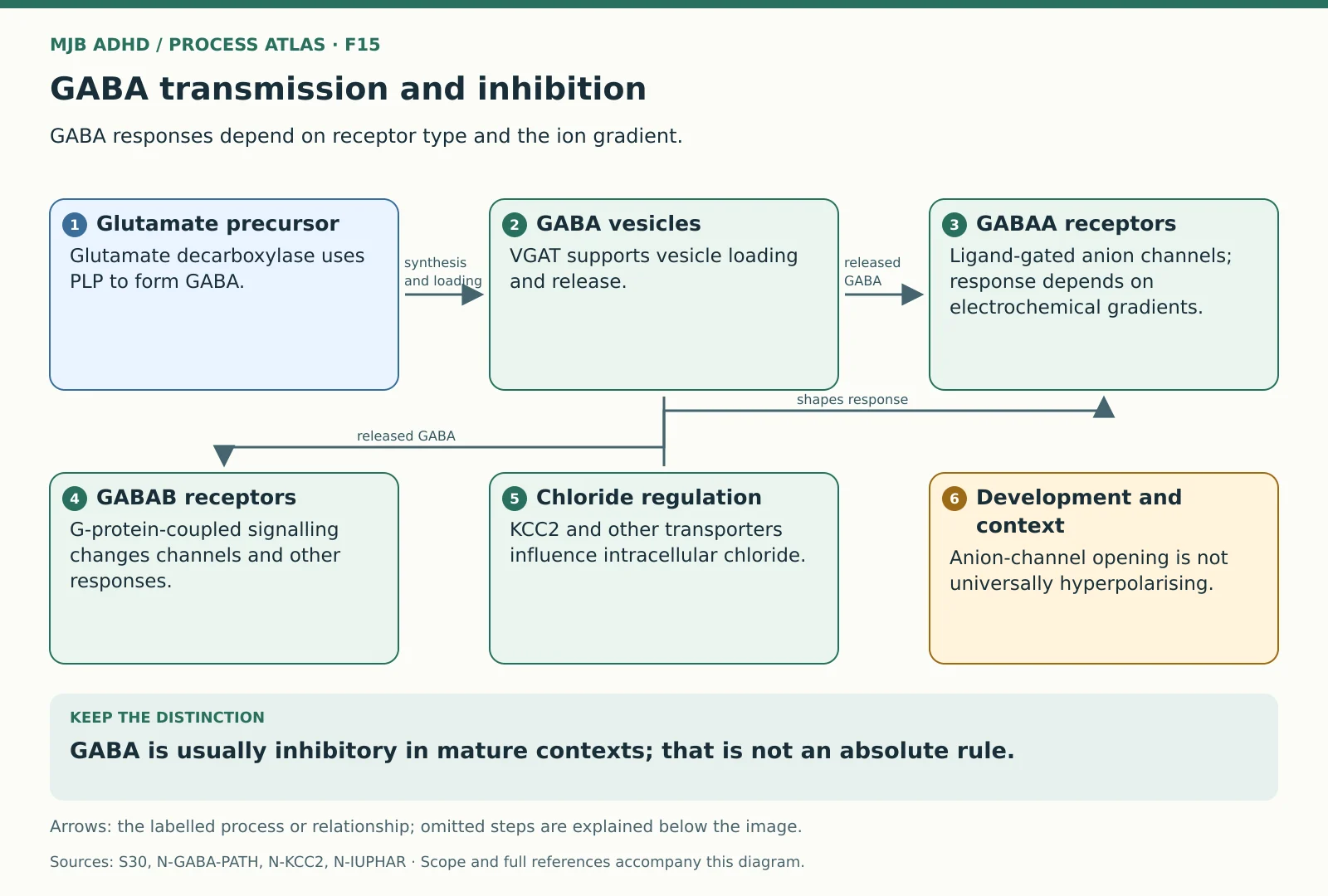 GABA transmission and inhibitionScoped educational mechanism
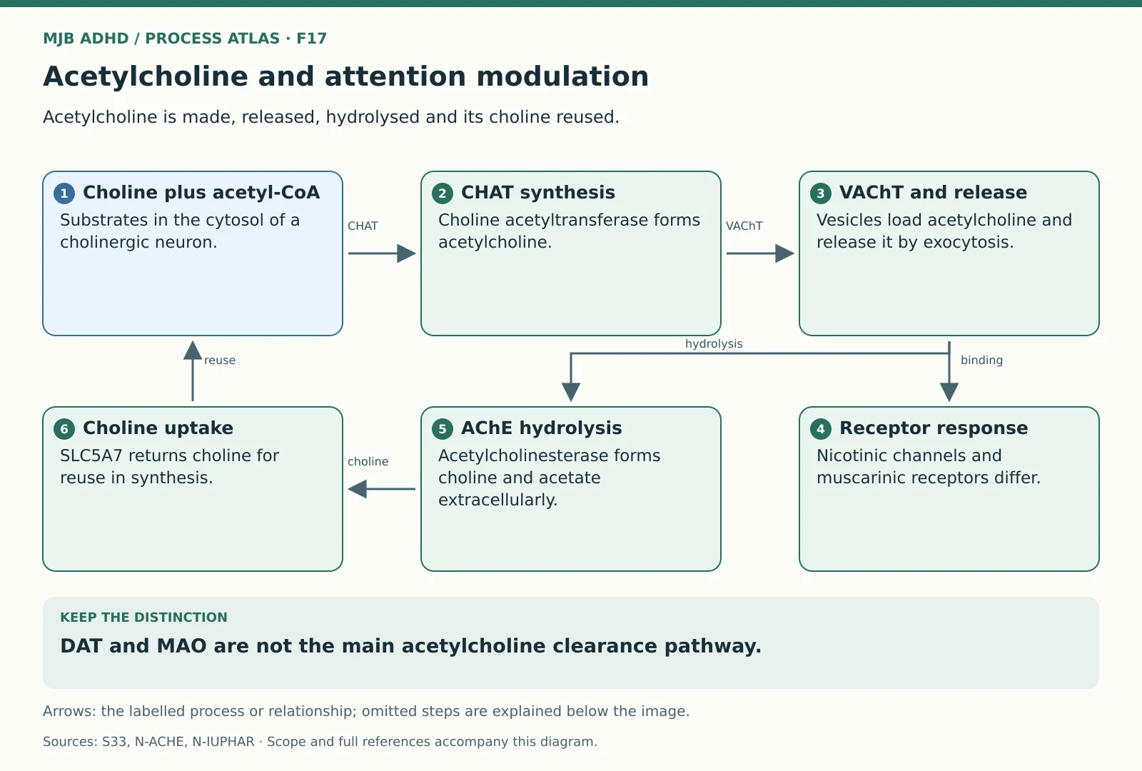 Acetylcholine and attention modulationScoped educational mechanism
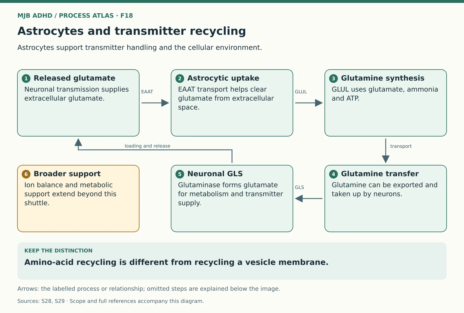 Astrocytes and transmitter recyclingScoped educational mechanism
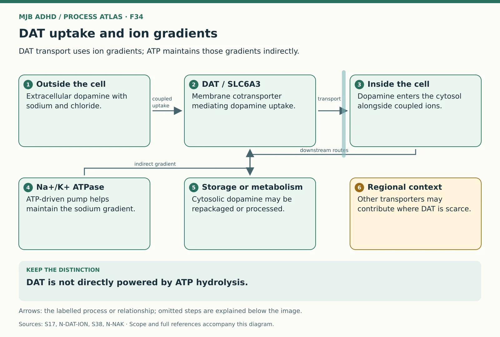 DAT uptake and ion gradientsScoped educational mechanism
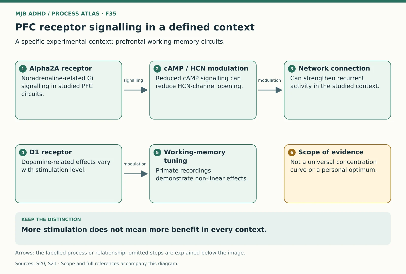 PFC receptor signalling in a defined contextScoped educational mechanism
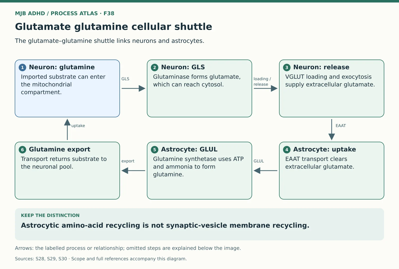 Glutamate glutamine cellular shuttleScoped educational mechanism
Energy and cellular support
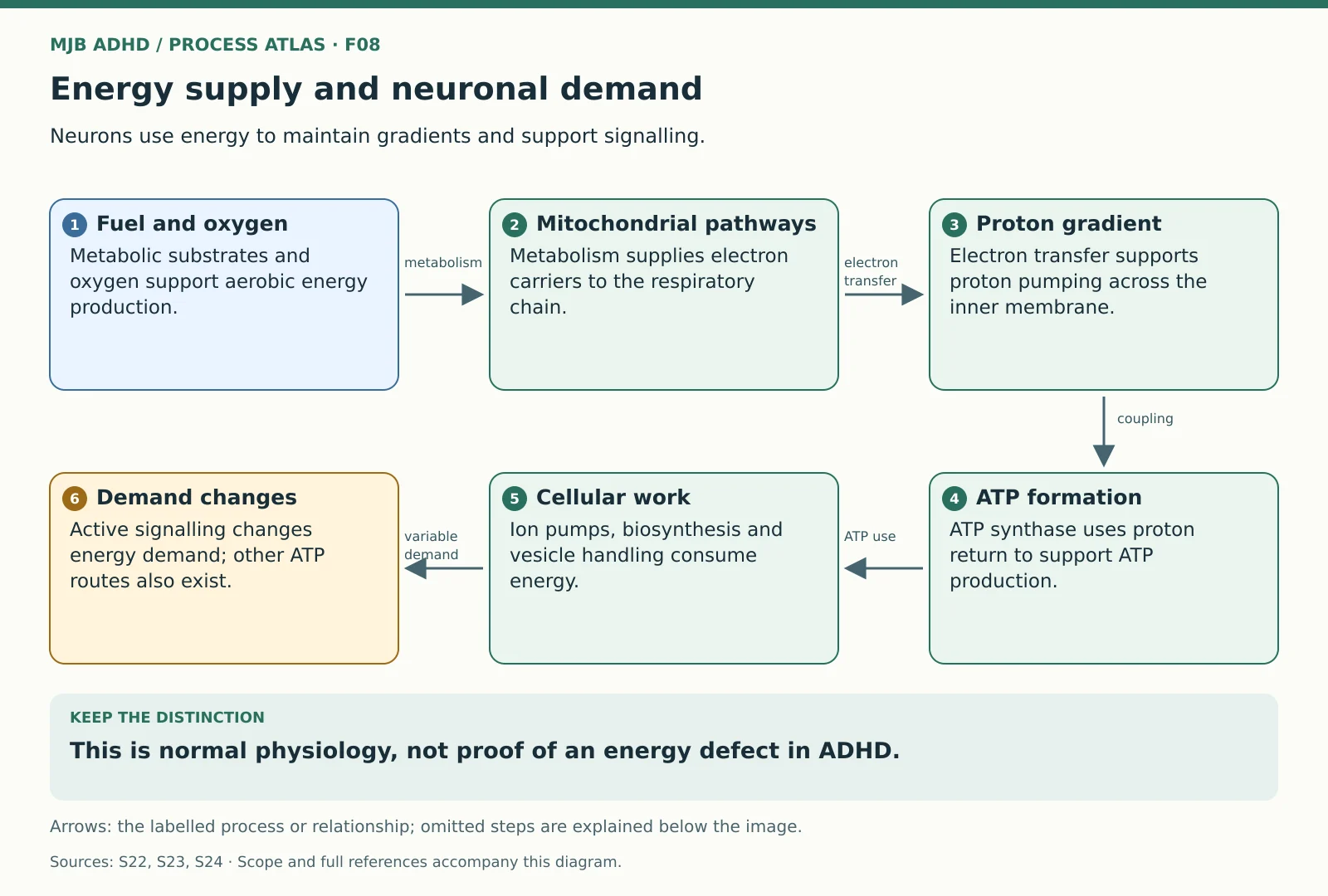 Energy supply and neuronal demandScoped educational mechanism
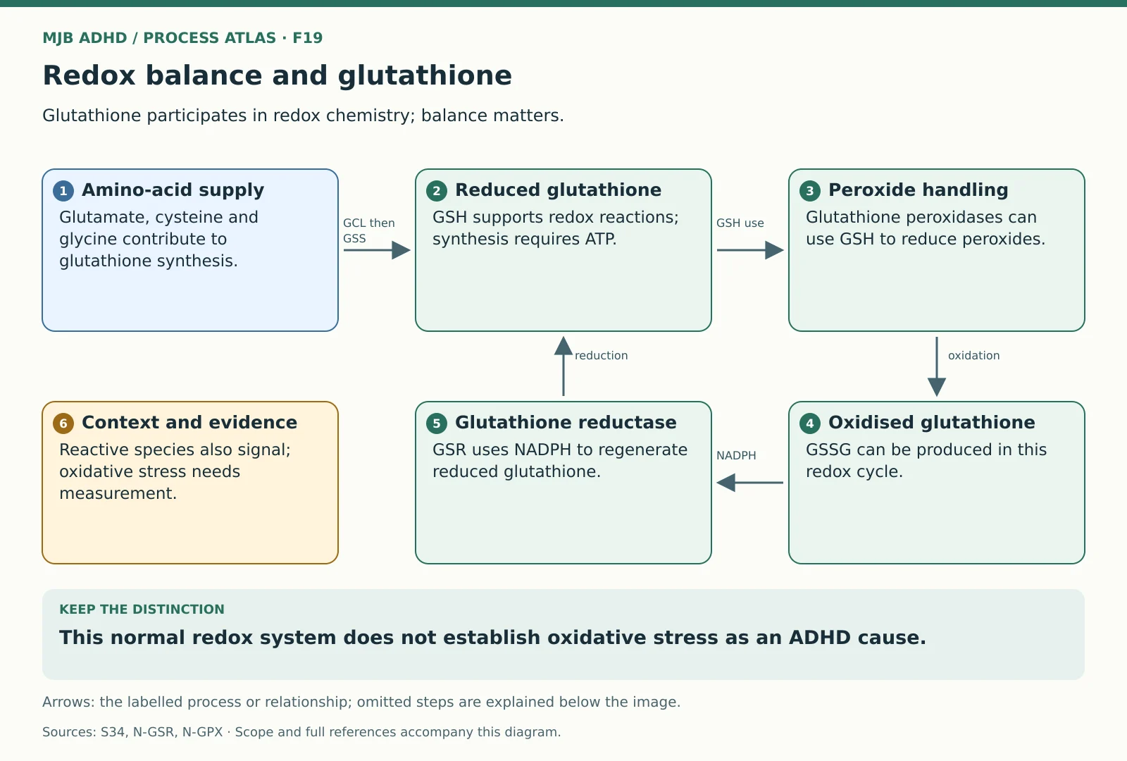 Redox balance and glutathioneScoped educational mechanism
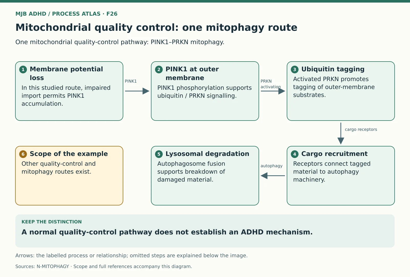 Mitochondrial quality control: one mitophagy routeScoped educational mechanism
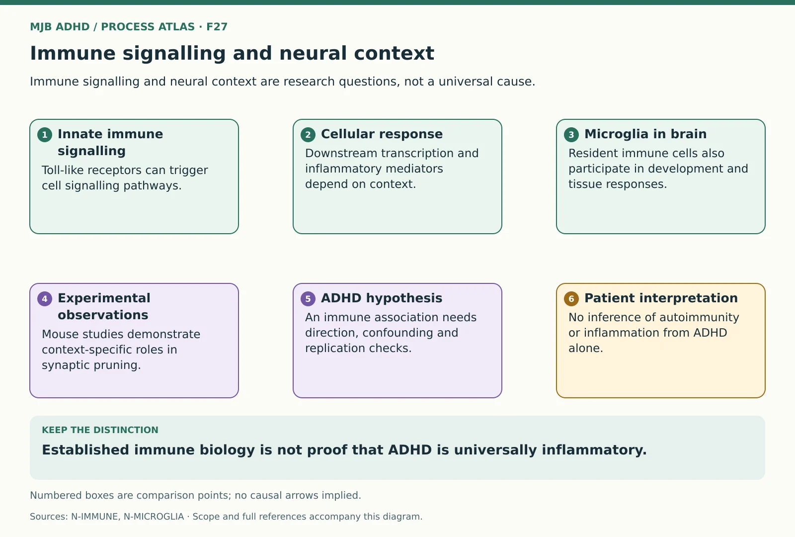 Immune signalling and neural contextEvidence interpretation
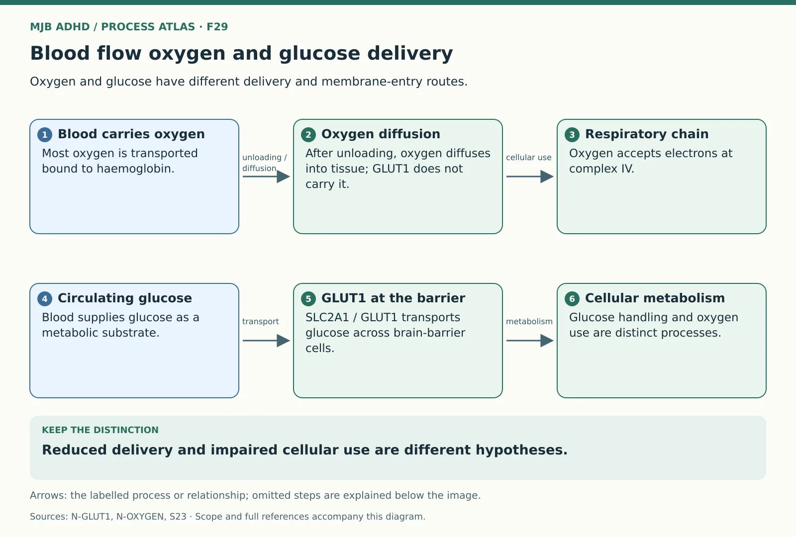 Blood flow oxygen and glucose deliveryScoped educational mechanism
 Mitochondrial membrane and respiratory chainEstablished cellular mechanism
Context and arousal
 Dopamine and noradrenaline projection systemsScoped educational mechanism
 Sleep circadian timing and arousalScoped educational mechanism
 Adrenal hormones: two tissue compartmentsScoped educational mechanism
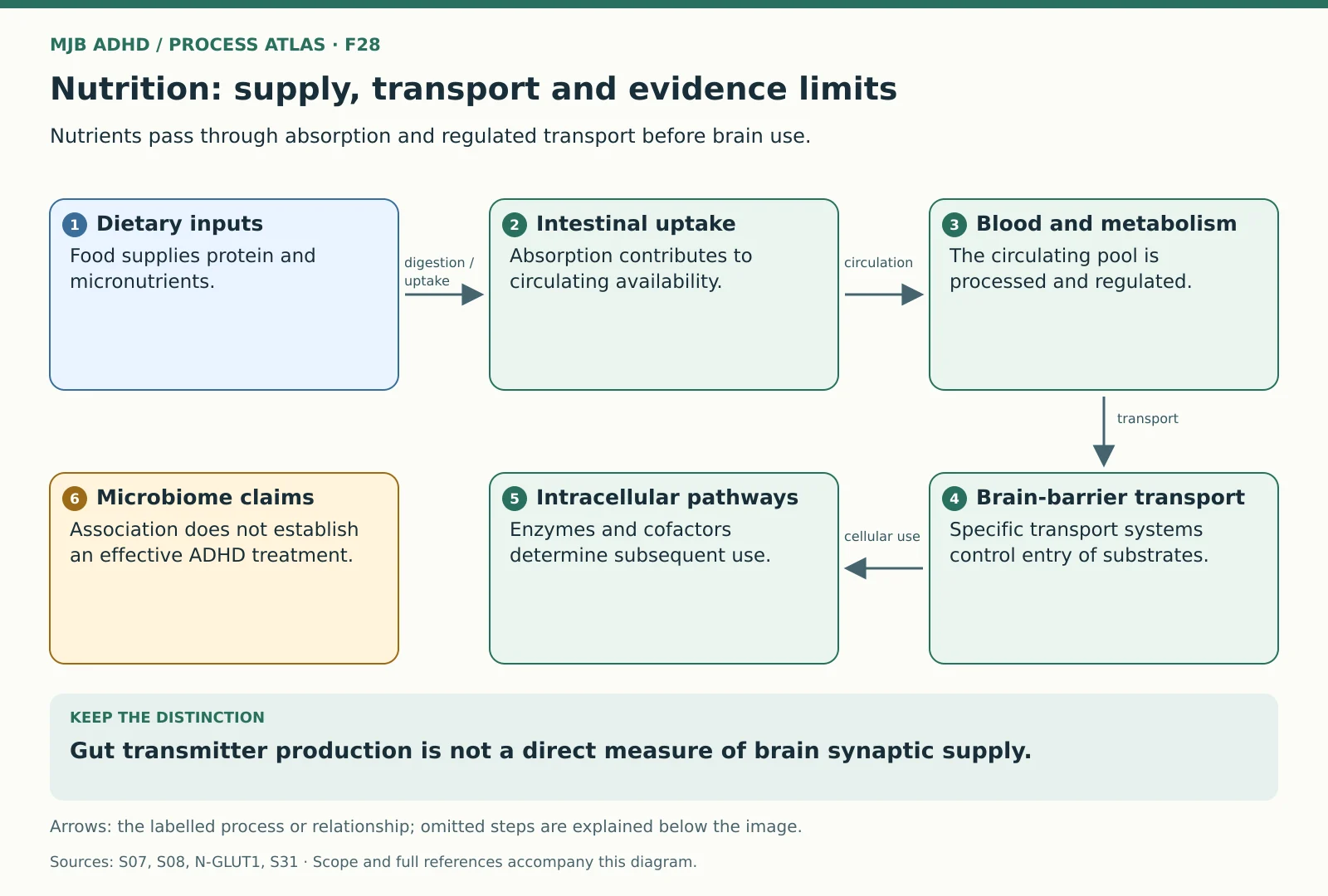 Nutrition: supply, transport and evidence limitsScoped educational mechanism
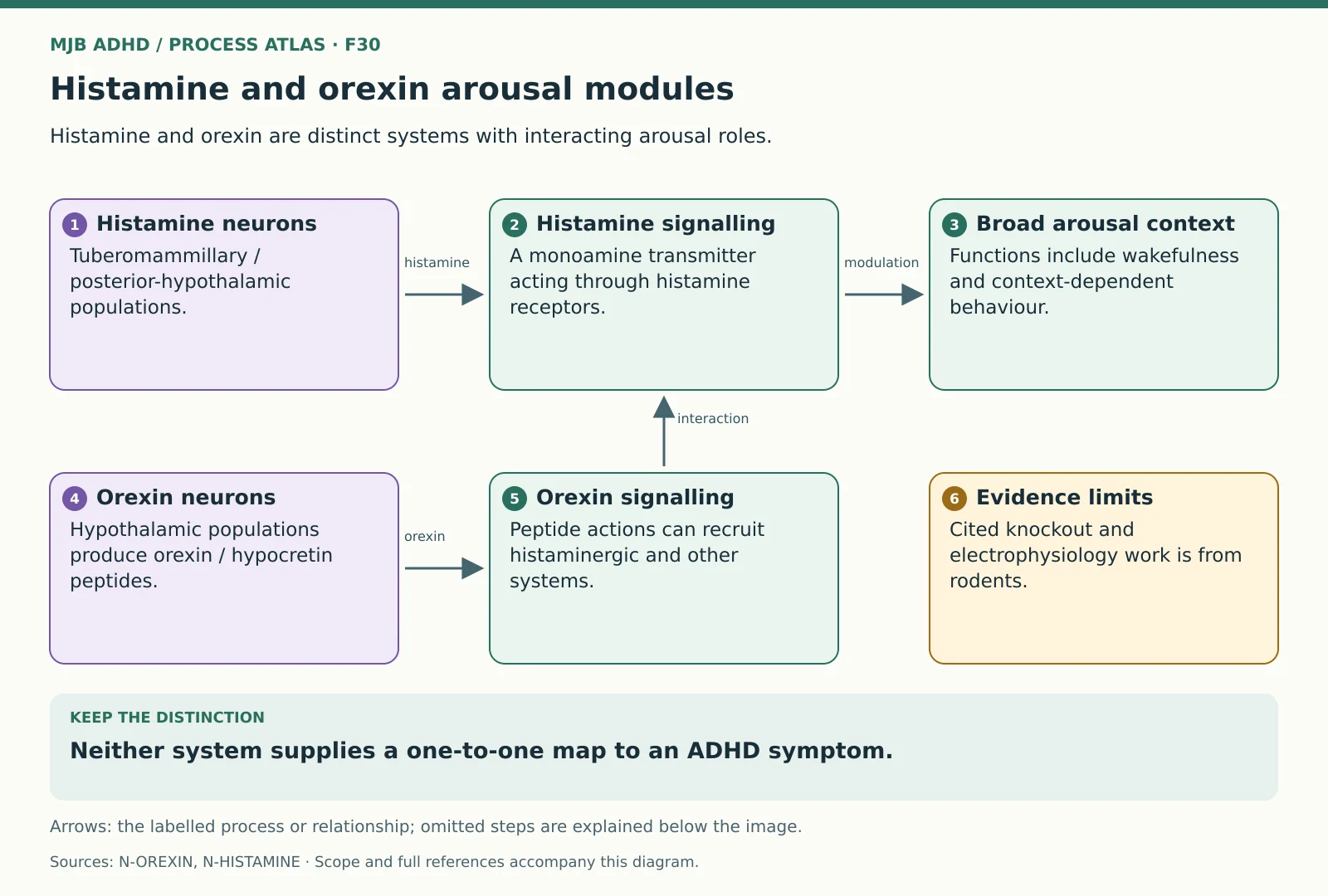 Histamine and orexin arousal modulesScoped educational mechanism
Medicine handling and targets
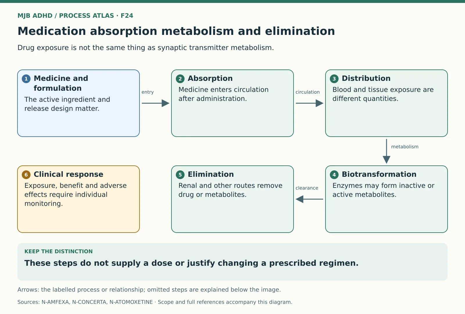
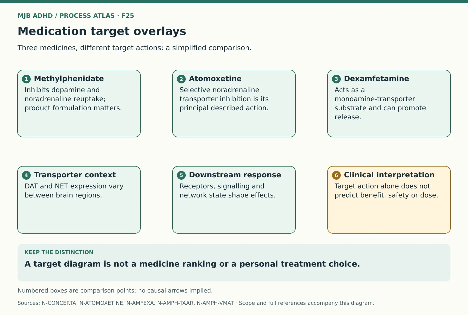
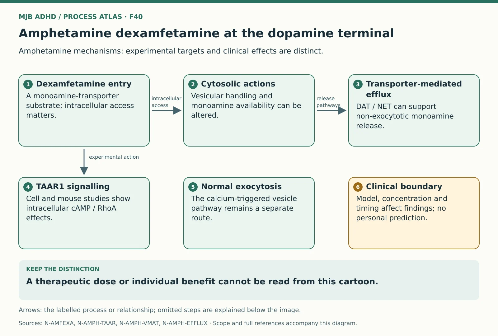
Four starting points
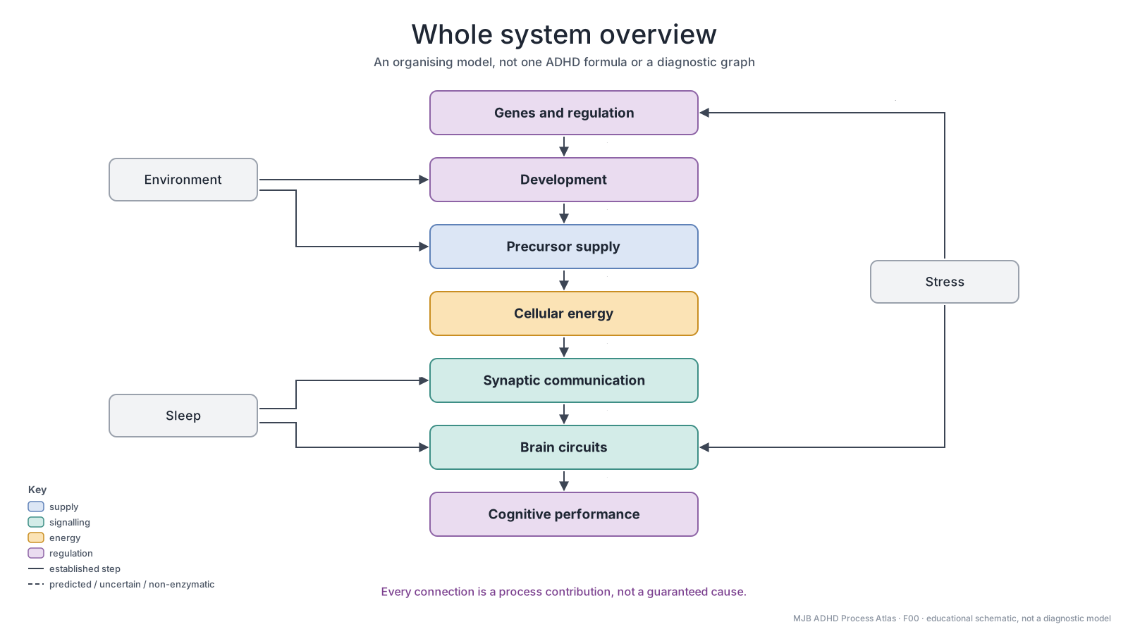
Whole system overview
An organising framework
Open larger diagram · Full-size PNG · Diagram page
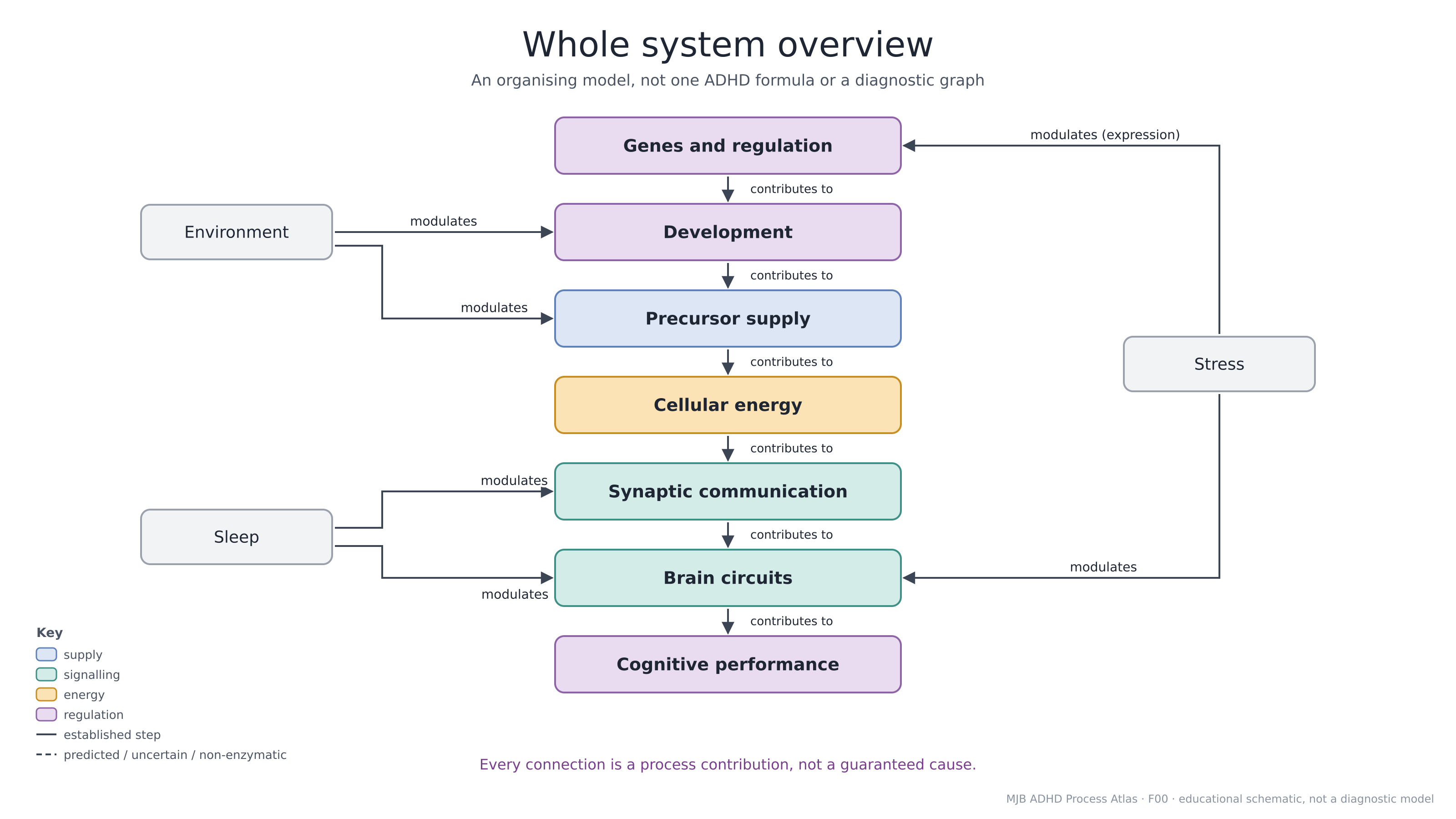
Read the diagram explanation and sources
Read from genes and regulation through development, precursor supply, cellular energy, synaptic communication and brain circuits to cognitive performance. Sleep, stress and environment are additional inputs. The connecting arrows organise relationships for learning; they do not demonstrate one causal chain, one defect shared by everybody with ADHD or a diagnostic test.
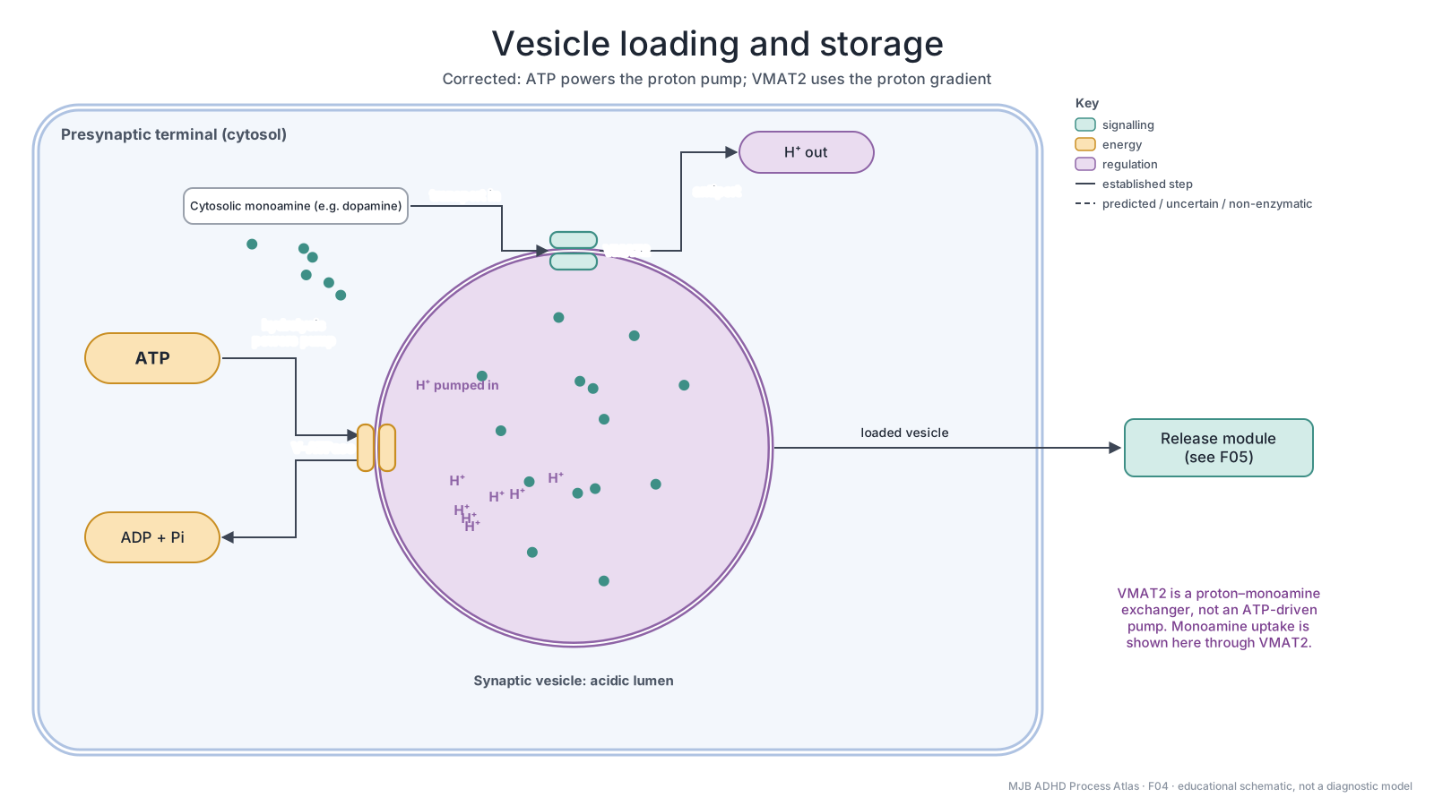
Vesicle loading and storage
Established transport mechanism
Open larger diagram · Full-size PNG · Diagram page
Read the diagram explanation and sources
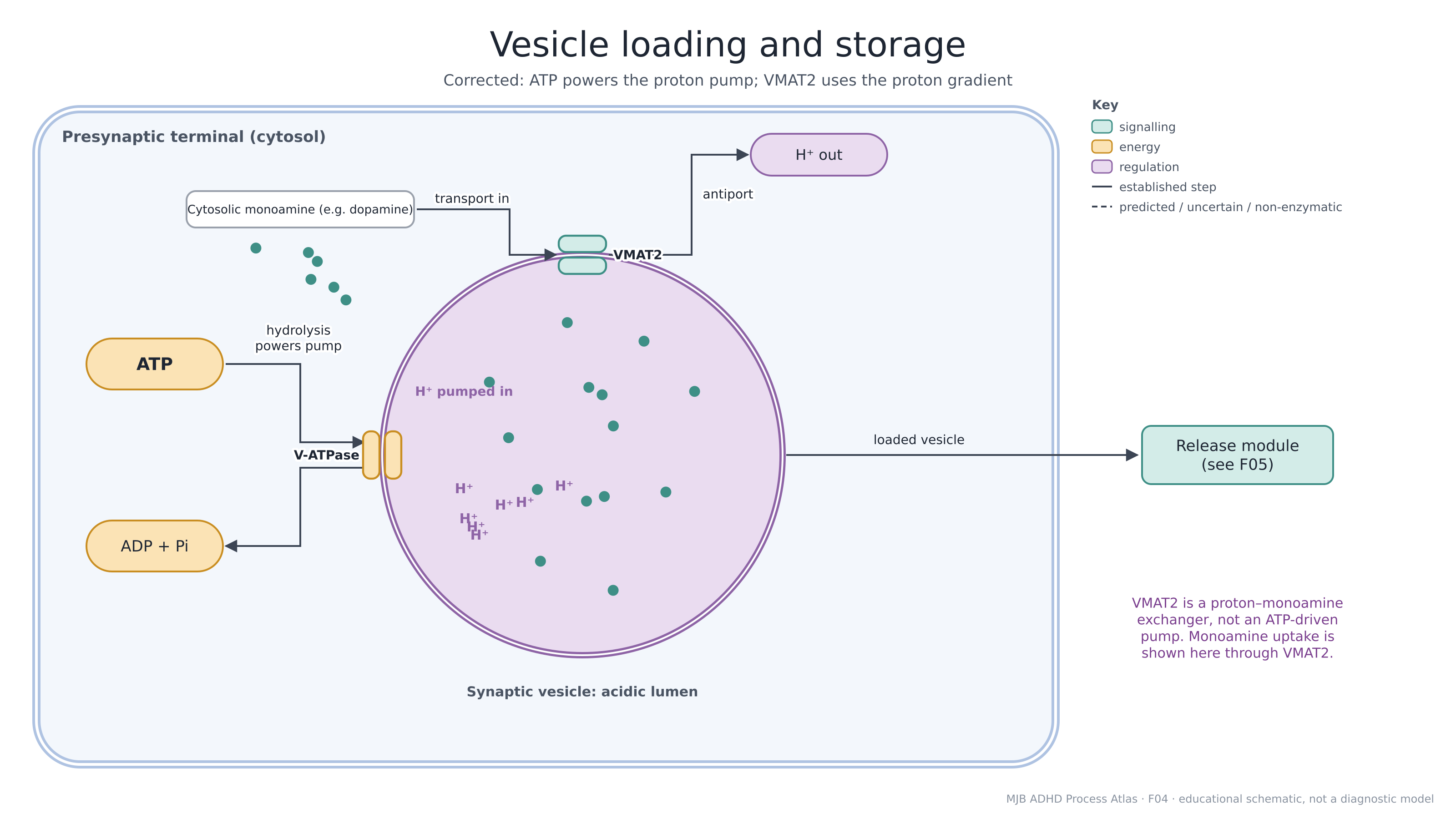
ATP hydrolysis powers the vesicular proton pump, V-ATPase, which acidifies the vesicle. VMAT2 exchanges vesicular protons for cytosolic monoamines such as dopamine. It therefore uses an existing proton gradient rather than directly hydrolysing ATP itself. Stored transmitter can subsequently be released by exocytosis. This explains a normal mechanism, not evidence of a VMAT2 defect in a particular reader.
Mitochondrial membrane and respiratory chain
Established cellular mechanism
Open larger diagram · Full-size PNG · Diagram page
Read the diagram explanation and sources
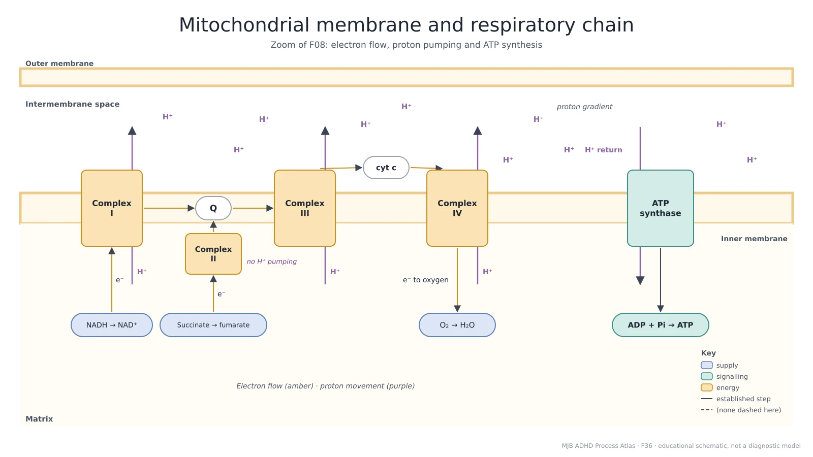
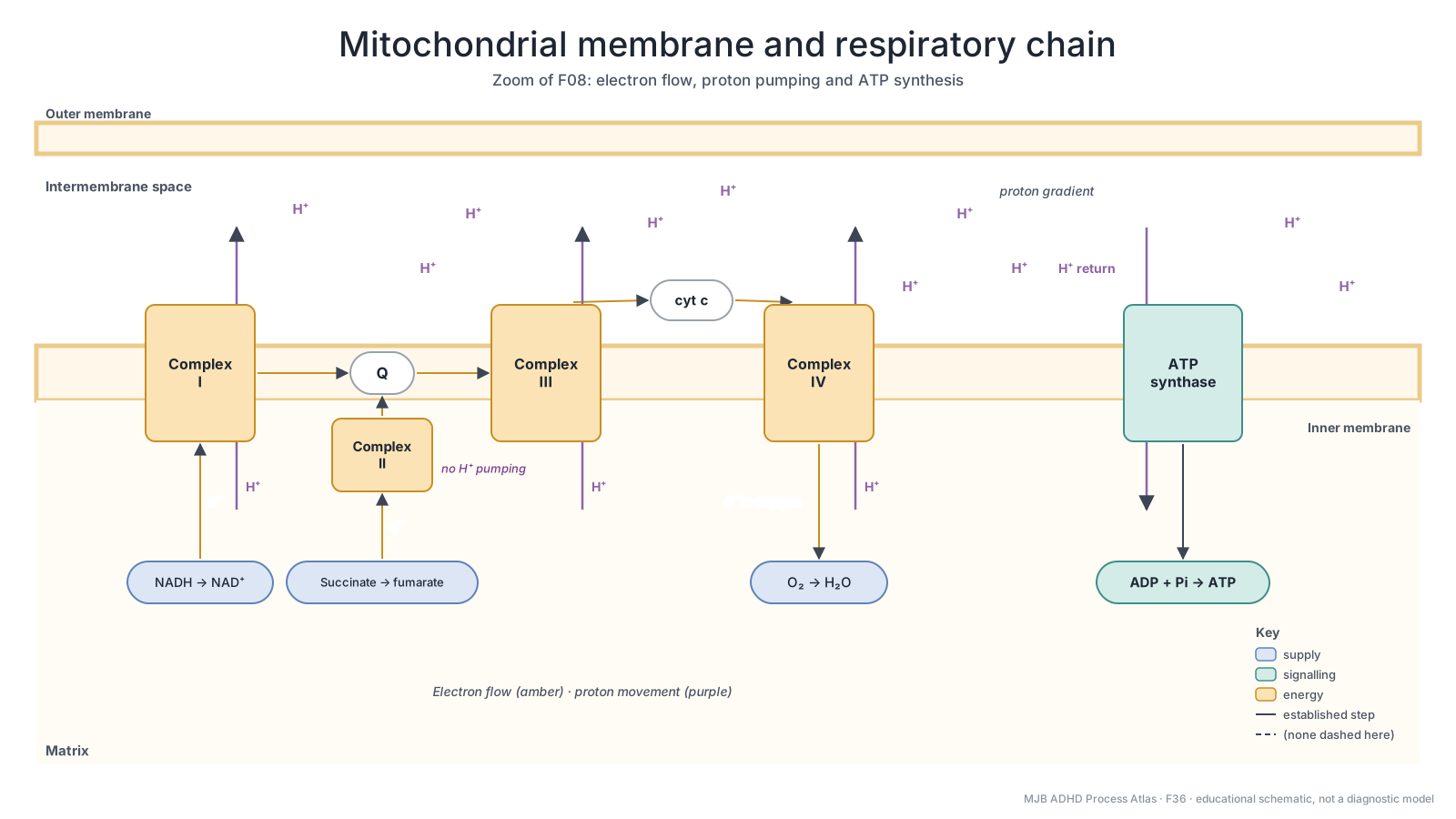
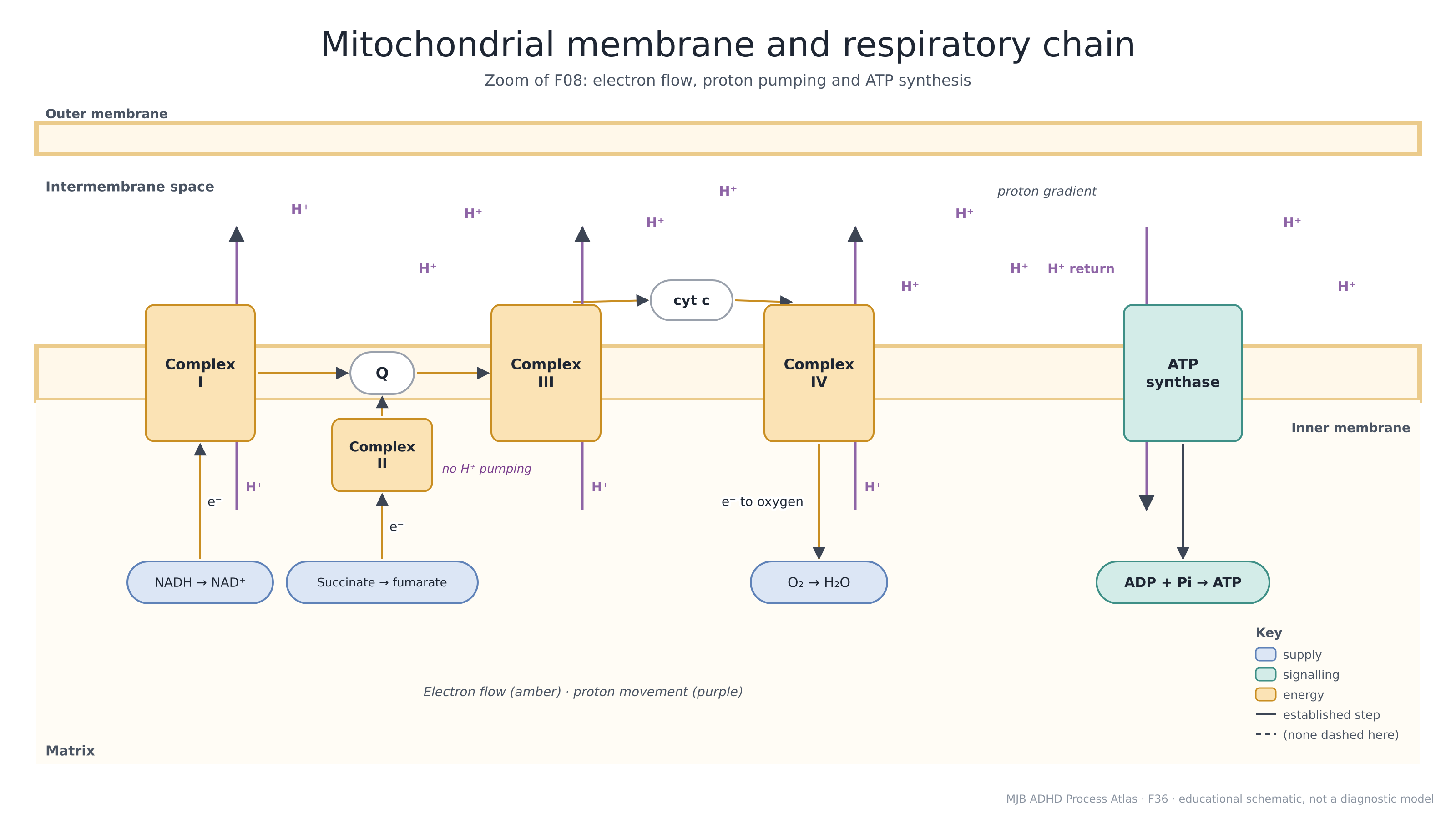
NADH supplies electrons through complex I, while succinate oxidation supplies electrons through complex II. Ubiquinone transfers electrons to complex III, cytochrome c carries them to complex IV, and oxygen is the final acceptor. Complexes I, III and IV pump protons from the matrix towards the intermembrane space. Complex II is membrane-bound but does not pump protons; its label is placed on the matrix side in this simplified drawing. Proton return through ATP synthase supports ATP formation from ADP and phosphate. The diagram does not establish mitochondrial dysfunction in ADHD.
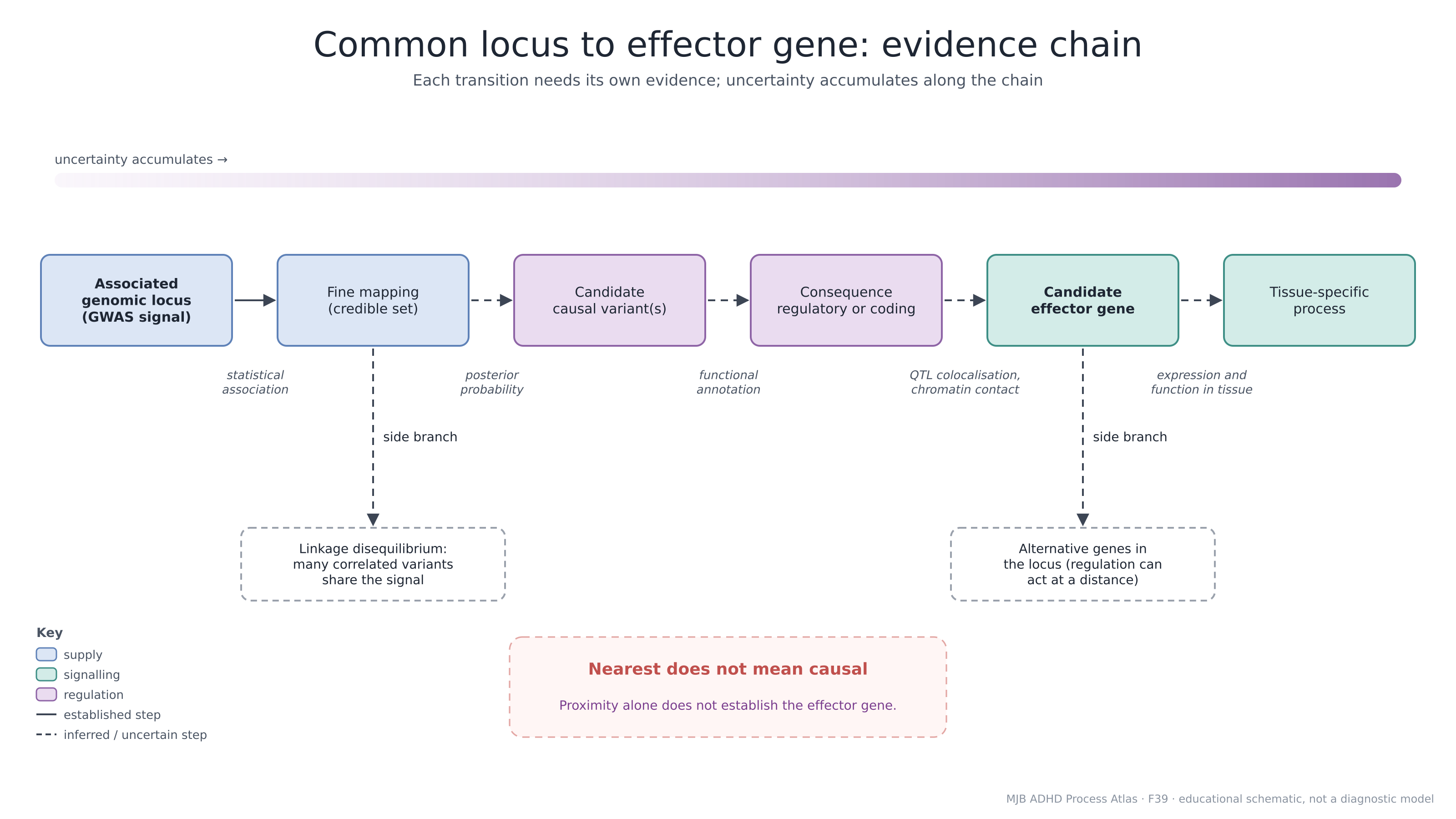
Common locus to effector gene evidence
Evidence interpretation framework
Open larger diagram · Full-size PNG · Diagram page
Read the diagram explanation and sources
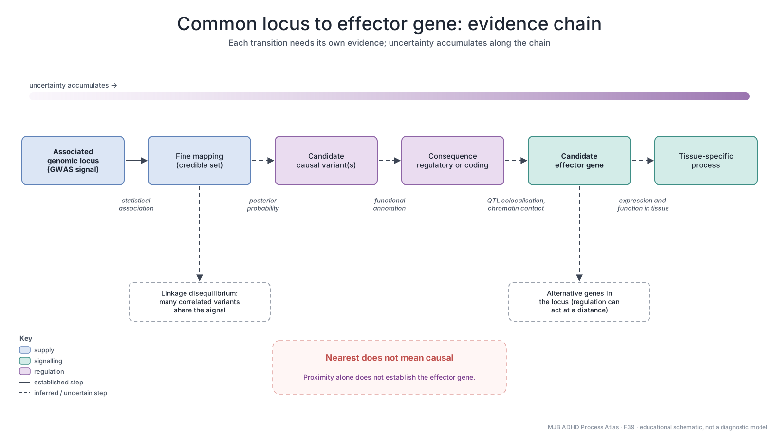
Begin with an associated locus, then evaluate fine-mapping, candidate variants, regulatory or coding consequences, potential effector genes and tissue-specific functions. Each step needs its own evidence. Linkage disequilibrium can leave several correlated candidates, and regulation may act over a distance. Proximity can be a clue but does not establish the causal gene by itself. These are complementary evidence types, not a mandatory sequence or a validated causal conclusion about one person.
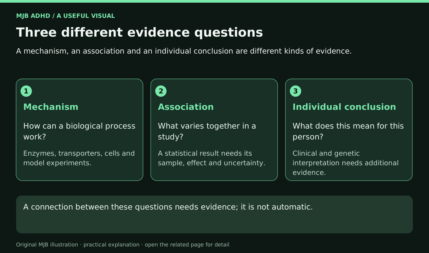
Three different evidence questions
Read the related explanation · Open full-size illustration
Read the image as text
A mechanism, an association and an individual conclusion are different kinds of evidence.
- Mechanism: How can a biological process work? Enzymes, transporters, cells and model experiments.
- Association: What varies together in a study? A statistical result needs its sample, effect and uncertainty.
- Individual conclusion: What does this mean for this person? Clinical and genetic interpretation needs additional evidence.
A connection between these questions needs evidence; it is not automatic.